Sensorimotor circuit for tactile perception
Our research interests reside mainly in the brain structures and functions that enable the sensory and motor control of our orofacial regions such as mastication, tasting or swallowing. Neural networks in our brain consist of numerous neurons and complicated connections among these neurons. That is one major reason why it is difficult to understand the mechanisms of our brain. We are trying to reveal “circuit diagrams” with respect to firing properties of neural elements at a single neuron level in the trigeminal sensory and motor system of rodents. Our animal studies use advanced anatomical techniques, single neuron recording/labeling, tissue clarification combined with electron microscopy (EM) and three-dimensional EM analysis. We believe that accumulated knowledge of network architecture in the trigeminal system would contribute to understanding mechanisms of nervous system in orofacial functions.
Central pattern generator for orofacial movement
We collaborated with an international research team to investigate the central pattern generator (CPG) regulating whisker movement (Moore et al., Nature, 2013; Matthews et al., J Comp Neurol. 2015; Deschenes et al., Neuron, 2016). We demonstrated that the CPG for whisking shares the neural circuit with the CPG for respiration.
Superior colliculus for vibrissal movement
We explored the neural circuit affecting the activity of the central pattern generator (CPG) for whisking. Kaneshige et al. (Front Neural Circuits, 2018) revealed that the superior colliculus (SC), which plays an important role in eye movement, also influences whisking, directly innervating the motoneurons in the facial nucleus for whisking muscles.
Movement regulation and axonal projection of the motor cortex
Shibata et al. (eNeuro, 2018) investigated the axonal projection pattern and the firing property of single neurons related to whisking in the motor cortex. This study revealed that the firing patterns of cortical neurons during whisking depend on pyramidal cell types in the motor cortex. Pyramidal tract (PT)-type cells projecting to the subcortical regions in the brain showed enhanced firing rate during a large-amplitude vibrissa movement, while callosally projecting intrateleencephalic (IT)-type cells preferentially exhibited a high firing rate during a small movement or a quiet state. This result indicates projection type-dependent coding of motor information.
Corticofugal projection modulates thalamic activity
Hirai et al. (Brain Struct Funct., 2018) investigated the effect of the corticofugal projection from the somatosensory cortex on the activity of the ventral posteromedial thalamic nucleus (VPM). This study demonstrated that cortico-thalamic input influences the resting membrane potential of VPM neurons, resulting in modulation of the firing mode and sensory gain of thalamic neurons. Thus, the cortical neurons can modulate the ascending sensory information via this cortico-thalamic innervation.
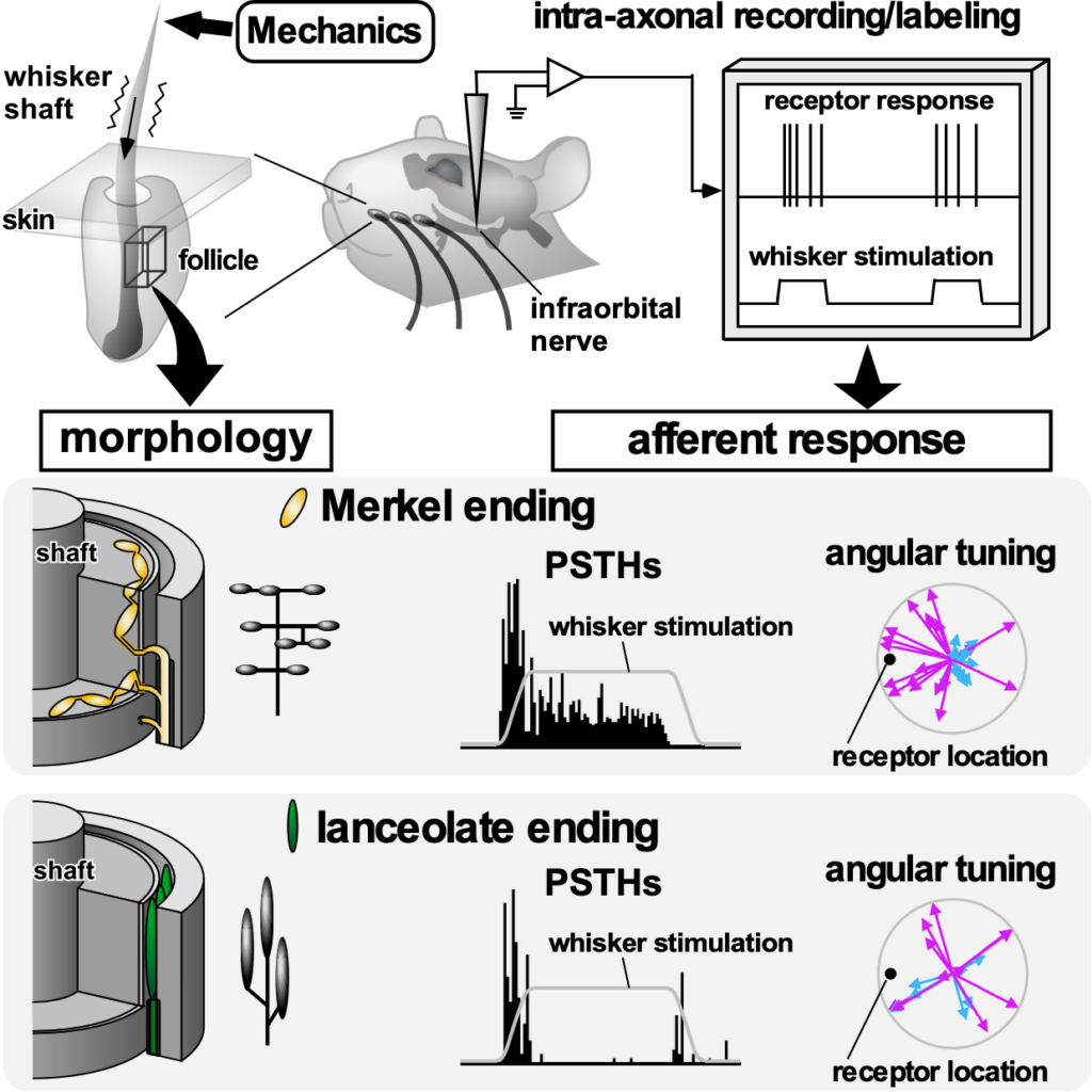
Response property of mechanoreceptors for tactile sensation
Mechanoreceptors for tactile sensation show unique response and morphological properties. Furuta et al. (Curr Biol., 2020) determined the relationship between morphological geometry of mechanoreceptors in whisker follicles and angular tuning to tactile stimuli. We are investigating how mechanical stimuli evoked by vibrissal movement shape neuronal responses based on anatomical properties of mechanoreceptors.
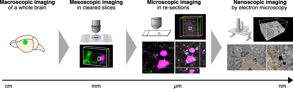
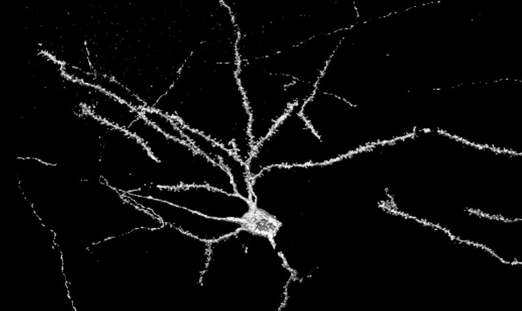
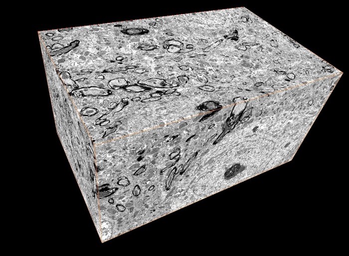
Development of zoom-in microscopy from the whole brain to nano-scale imaging
Our research team developed a seamless imaging technique from a whole brain to nano-scale imaging using a tissue clearing method (Furuta et al., iScience, 2021). This strategy allows multi-scale imaging by coupling light and electron microscopy with preservation of microstructure of the brain tissue.